Friday, August 29, 2014

The Eyes Have It: The Drosophila eye as a model for understanding gene expression regulation


Male a Female Fruit Flies.  Cartoon Via WikiHow
Those tiny little fruit flies that swarm when you pick up a past-its-peak banana at home will almost certainly have red eyes.  In the lab however, fruit fly eyes come in many different varieties.  There are many genes involved in the production of pigments responsible for the eye color in flies and these mutants have been essential to the study of genetic inheritance of traits since the earliest days of fruit fly genetics and Thomas Hunt Morgan. 
Figure ©1965 by Srb et a.;. all text material ©2006 by Steven M. Carr
Mutant flies have been historically named for their phenotypes, how they changed the appearance or behavior of flies.  The white mutation was eventually traced to a gene that is also named white, in honor of its associated mutant phenotype.  This explains the somewhat confusing fact that a functional white gene is necessary for flies to have red eyes (non-functional white gene as found in the white mutant results in white eyes).  This white gene has provided an extremely useful tool for understanding chromatin regulation.  By inserting a functional white gene in different regions of the genome, the ability of the white gene to be expressed can be determined by the appearance of the eyes of the fly.  When the white gene is in a region of the genome with open and accessible chromatin, allowing gene expression, the fly’s eyes are red.  If the white gene is in heterochromatic region, where the gene is inaccessible to transcription factors and the gene expression machinery, the eyes of the fly show variegated red pigment.  Some of the ommatidia, the individual units that make up the compound eye of the fly, will be red, while others will be white, indicating that the gene can be expressed at sometimes but not others due to heterochromatin formation.  This biological representation of gene expression is known as position effect variegation (PEV).
From left to right:  Red eyed, wild-type fly eye; Fly with non-functional white gene; Fly eye exhibiting position effect variegation  (PEV).  
We make use of this system to understand how genome location and DNA sequence can affect how genes are expressed.  The fourth chromosome of Drosophila melanogaster is largely heterochromatic, highly condensed, and yet the genes that found there can be expressed.  What is about those genes that allow them to escape the repressive effects of heterochromatin?  This is the question we are trying to address with the project I will be discussing here. 


Sunday, February 9, 2014

Packing it in: DNA and its place in the nucleus


Attribution: Darryl Leja, NHGRI.


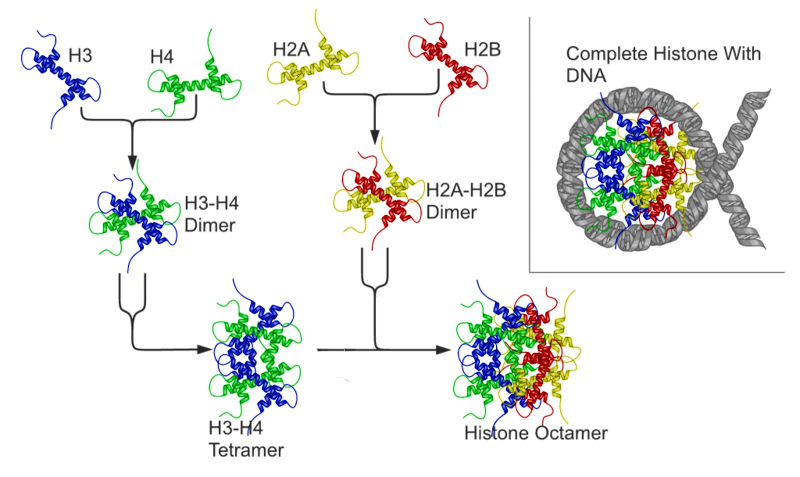
More than just the hereditary material, DNA is in constant use within cells.  DNA is a dynamic substance; the quintessential double-helix is in constant and intimate connection with other molecules, namely protein complexes known as nucleosomes that are composed of histones.  DNA coils around histones, which in turn coil and form higher-order structures as well.  This all creates a complicated three-dimensional environment in which DNA is not easily accessible at all times. 


The Nucleosome.  Attribution: Richard Wheeler
[CC-BY-SA-3.0 or GFDL], via Wikimedia Commons.

This combination of DNA and associated proteins is known as chromatin and it is chromatin that allows for fine-tuned regulation of gene expression.  Both DNA and histones in chromatin are the targets of modifications.  The addition of chemical groups, such as methyl moieties, changes the properties and accessibility of DNA within chromatin.  As not every gene is expressed at all times and in all tissues, the complex combination of chemical modifications in chromatin sets up the mechanism by which gene expression is regulated, this often called the chromatin code.   By adding methyl or acetyl groups to the ends of histone molecules, the so-called histone tails, the degree to which those tails can physically interact with nearby DNA is altered.  This creates two fundamental types of chromatin: euchromatin and heterochromatin.  In a euchromatic state, histone molecules are associated with DNA but not so tightly as to preclude the molecular machinery responsible for gene expression from accessing the DNA, thereby allowing for gene expression and RNA production.  In contrast, heterochromatin is very tightly packaged and dense, and therefore genes associated with heterochromatin are thought to be blocked from expression.  

There are many great resources available regarding DNA and chromatin and its function in biology, including the ModEncode EducationalSupplement. 

Friday, January 17, 2014

Welcome: Let's Introduce Heterochromatin

Hello and welcome!  This blog is dedicated to explaining the role of heterochromatin in gene expression and giving a glimpse into the real world practice of scientific research in the laboratory.  We will focus most specifically on heterochromatin in Drosophila melanogaster (fruit flies), as it is the model organism we utilize in our research.  This work is taking place in the Elgin Laboratory at Washington University in St. Louis, and is part of a project funded by the National Science Foundation.  
The posts in this blog will be aimed at educating people about the function and regulation of heterochromatin, focusing on its importance in biological processes.  A basic knowledge of genetics will be helpful in understanding the subjects covered here, but every attempt will be made to make these posts accessible to non-experts.
This blog will be updated weekly, starting with a series of informative posts discussing heterochromatin, gene expression, and flies.  Bookmark this page and join us next week for a definition and discussion of heterochromatin.